Diagnosing Keratoconus

Diagnosing Keratoconus and Treatment Options

The goal of treatment for iLink® corneal cross-linking patients is to slow or halt the progression of keratoconus. For these patients, continued progression often results in loss of visual acuity or decreased tolerance to contact lens wear, caused by the ongoing changes in the cornea. This means early diagnosis is critical so that progressive keratoconus can be treated sooner.1

Bring advanced topography into your practice. Glaukos and Topcon have partnered to offer optometrists the advanced technology of Topcon CA-800 topographer at a lower cost of entry.

[Gelles2017/p1/col1/para2/lines6-9; col2/para1/lines5-8; p3/col1/para2/lines1-7]

Left untreated, 1 in 5 patients with progressive keratoconus may require a corneal transplant. More than half of these patients could need multiple transplants within 20 years. 2 3

[Pramanik] [Maharana]

Explore information about iLink®

Learn More

Identifying keratoconus patients

Appropriate patients who have been diagnosed with progressive keratoconus or corneal ectasia following refractive surgery may be candidates for iLink® corneal cross-linking.

  • Keratoconus is a bilateral, progressive corneal ectasia resulting in irregular astigmatism and loss of visual function, with onset in teenage years.
  • Corneal ectasia, a non-inflammatory condition marked by progressive corneal steepening and thinning, is a rare but serious complication of vision correction procedures.

What are the signs and symptoms of progressive keratoconus?

Early signs of keratoconus may include asymmetric refractive error, high or progressive astigmatism, or reduced best corrected visual acuity. The onset of keratoconus often occurs in the teenage years or early twenties but can start at any time.

Patient symptoms may include:

  • Constantly and regularly changing refractive errors
    Blurry vision
  • Increased light sensitivity
  • Difficulty driving at night
  • A halo around lights and ghosting (especially at night)
  • Eye strain
  • Headaches and general eye pain
  • Eye irritation, excessive eye rubbing

Keratoconus, especially in the early stages, can be difficult to diagnose, and all of the above symptoms could be associated with other eye problems.

What tools do I need to observe the signs of keratoconus?

Keratometry
  • Fleischer’s ring
  • Vogt’s striae
  • Apical scarring
Corneal topography or tomography
  • Topography and corneal thinning
Corneal OCT
  • Topography and corneal thinning

Topography Examples
Patient with Keratoconus.
Patient with normal astigmatism.

The iDetect KC Program

iDetect KC helps equip optometrists with advanced topography so they can begin integrating the 3 pillars of modern keratoconus care into their practice.

Advanced topography enables earlier diagnosis and monitoring to help preserve patients’ vision by intervening sooner
Optometrists can then refer patients to an ophthalmologist who can
perform iLink®
With topography, optometrists can provide long-term, continued medical care for patients while supporting the expansion of their contact lens service

For more information on bringing an advanced topographer into your optometry practice…

Contact Us

Collaborating with surgeons and post-op care

iLink® corneal cross-linking provides the opportunity for collaborative care between optometrists and ophthalmologists.

Optometrists are an essential part of the ongoing medical care keratoconus patients receive after they have been treated with iLink® corneal cross-linking. It may be necessary for an optometrist to increase the frequency of a patient’s monitoring exams or fit them with contact lenses several times while their corneas stabilize.

Epithelial healing

Post-operative care management is up to the physician’s discretion. Although there is certainly some variation, corneal specialists will often see patients for the 1-day and 1-week visits and then, once the epithelium is intact, send them back to co-managing doctors for the remainder of the follow-up care. Patients should be monitored for resolution of epithelial defects.

Contact lens fitting

Scleral lenses or other vaulted designs are a good option for keratoconus patients as they do not rest on the corneal surface and avoid disrupting the epithelial healing. It is important to make patients aware that vision may change slightly during the first few months, and that the contact lens prescription may need to be updated.

For contact lens fitting purposes, one can expect that the corneal curvature may change after cross-linking, but not dramatically. It is not uncommon to notice an initial corneal steepening followed by flattening in the first months after an iLink® procedure. More frequent contact lens evaluations may be required in the first year after the procedure while the cornea stabilizes.

Clinical notes

“We report good visual results in patients who were fit in contact lenses after having undergone corneal cross-linking. Contact lens fits were as early as two weeks (soft lenses), but ranged depending on patient’s functioning needs and observed changes in corneal topography in the postoperative healing period. Gas permeable lenses were fit at the earliest 3 months postoperatively. Contact lens choice is a balance between patient’s needs and the lens able to achieve good comfort with optimal visual results.*”

-Clark Chang, OD, MS

*Results may vary

Resources for diagnosing keratoconus

Please Select Your Country

Continue

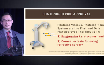
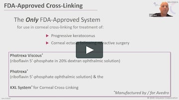
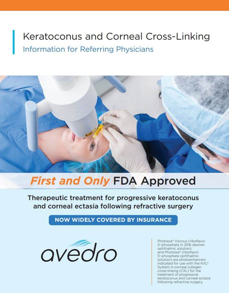
Using Photrexa® Viscous (riboflavin 5’-phosphate in 20% dextran ophthalmic solution), Photrexa® (riboflavin 5’-phosphate ophthalmic solution), and the KXL® System, the iLink® corneal cross-linking procedure from Glaukos is the only FDA-approved therapeutic treatment for patients with progressive keratoconus and corneal ectasia following refractive surgery.*1

[Photrexa IFU/p1/col1/para3/lines1-4]

Important Safety Information

Corneal collagen cross-linking should not be performed on pregnant women.

Ulcerative keratitis can occur. Patients should be monitored for resolution of epithelial defects. The most common ocular adverse reaction was corneal opacity (haze). Other ocular side effects include punctate keratitis, corneal striae, dry eye, corneal epithelium defect, eye pain, light sensitivity, reduced visual acuity, and blurred vision.

These are not all the side effects of the corneal collagen cross-linking treatment. For more information, go to Prescribing Info to obtain the FDA-approved product labeling.

You are encouraged to report all side effects to the FDA. Visit www.fda.gov/medwatch, or call 1-800-FDA-1088.

*Photrexa® Viscous and Photrexa® are manufactured for Avedro. The KXL® System is manufactured by Avedro. Avedro is a wholly owned subsidiary of Glaukos Corporation.

Indications

Photrexa® Viscous (riboflavin 5’-phosphate in 20% dextran ophthalmic solution) and Photrexa® (riboflavin 5’-phosphate ophthalmic solution) are indicated for use with the KXL System in corneal collagen cross-linking for the treatment of progressive keratoconus and corneal ectasia following refractive surgery.

Reference

1. Photrexa Prescribing Information. Burlington, MA: Avedro, a Glaukos company 2022